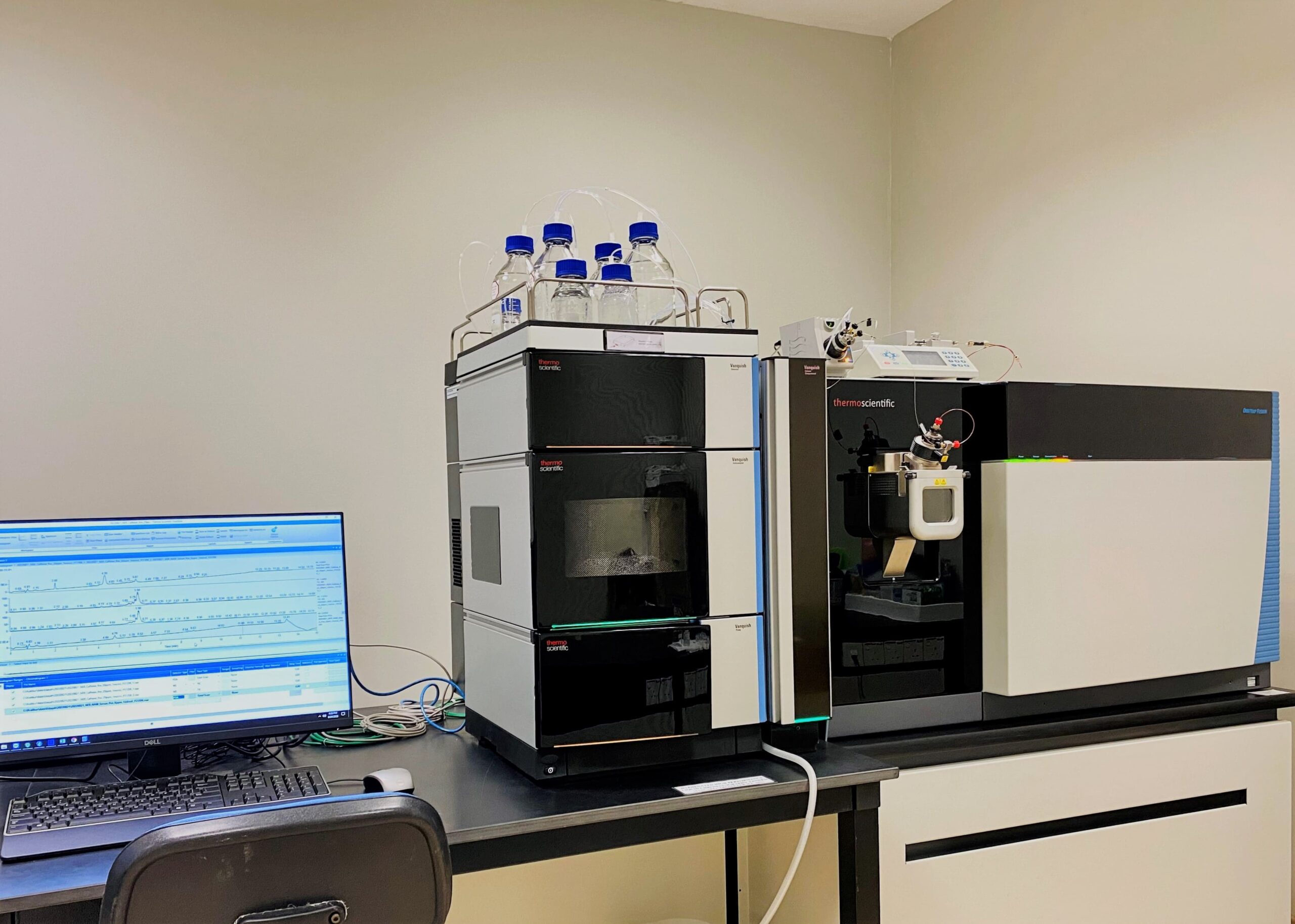
Liquid chromatography-mass spectrometry (LC-MS) is increasingly becoming a preferred technique for liquid chromatography. LC-MS bioanalysis combines liquid chromatography’s resolving power with the detection capacities of mass spectrometry. During LC-MS sample analysis, the liquid chromatography component separates the sample and introduces individual sample components into the mass spectrometer for detection. The mass spectrometer detects charged ions to identify individual sample components. LC-MS sample analysis provides data on molecular weight, identity, structure, and quantity of sample components. The current article discusses the working of LC-MS assays. Similar to toxicokinetic TK analysis, LC-MS bioanalysis requires robust LC-MS method development and validation initiatives.
Working of LC-MS mass spectrometry
Liquid chromatography physically separates the analytes present in the study sample. The sample solution is introduced into the mobile phase of the liquid chromatography. This mobile phase is pumped through the stationary phase that is made up of a column generally filled with silica particles. As the mobile phase and sample solution mixture reach the stationary phase, the mixture component interacts differently with the stationary phase depending upon their physical and chemical properties. Based on the interaction between the stationary phase and sample components, liquid chromatography separates analytes through different modes, including partition ion exchange, size exclusion, and affinity chromatography.
Today, multiple detection systems are available and coupled with liquid chromatography for analyzing different samples. However, the mass spectrometer is a sensitive, selective, and universal detecting system.
The eluent with separated analytes coming out of liquid chromatography is not allowed to enter the mass spectrometer directly. The primary reason for this limitation is that liquid chromatography operates at ambient pressure, while mass spectrometers operate under a vacuum. Hence, both these systems are joined through an interface, where the elements enter this interface, and heat is applied to evaporate the sample and vaporize and ionize the analyte molecule. This step is critical as MS detectors can only measure gas phase ions.
Must Read: ELISA Immunoassays: An Essential Tool In Biomedical Research
After entering the mass spectrometer unit, the analyte ions are subjected to magnetic and electric fields, resulting in different flight paths. Varying intensities of magnetic and electric fields ensure the separation of analyte ions, which are then measured based on the mass-to-charge value. The ions determined during LC-MS analysis are plotted where the peak intensity is plotted against the retention times. Each chromatogram point correlates to a mass spectrum. The compound mass spectrum provides data about the mass of the drug compound and helps determine the compound structure based on the isotopic mass peaks.
Researchers operate mass spectrometers through two modes: scan mode and selected ion monitoring mode. When operating in the scan mode, the researcher sets the settings to identify ions from low to high mass to charge value within a predefined period. Scan mode is ideal for analyzing unknown samples or when enough information is not available for ions in a sample. On the other hand, SIM mode measures specific mass to charge values and is preferred for quantifying known compounds in study samples.
In Conclusion
LC-MS systems are becoming mainstream due to their sensitivity, selectivity, and reliability. Besides, alternatives such as tandem mass spectrometry have further skyrocketed the applications of LC-MS systems in drug discovery and development studies.


